baby chest x ray technique
In the PA view the patient stands with the shoulders rotated anteriorly and depressed hands on the hips. Reproduction of the chest must extend from the cervical trachea to T12L1.

The Forbidden Chest X Ray Tension Pyopneumothorax The American Journal Of Emergency Medicine
These tests expose children to low doses of radiation.

. 10 It is unclear whether rates have decreased over time. X-rays are a kind of imaging test. The patient will place their chest against a plate which digitally records the image.
Pediatric Chest Screen 70-80 DIGITAL OPTIMUM kVp Universal CR Technique Chart using a standard 21 LgM Part View kV mAs kV mAs kV mAs Abdomen AP Grid 85 10 -15 85 20 - 25 85 30 - 40 Ankle AP 70 18 70 2 70 25 Ankle Obl 70 16 70 18 70 22 Ankle Lat 70 15 70 16 70 2 Chest -Adult AP 400 - tt -72 85 2 - 25 85 32 - 4 90 5 - 64. CHEST Body Part Grid mAs CM kVp PA Chest Y 4 14-15 85 2 20-21 100 6 26-27 110 72 6 16-17 85 3 22-23 100 9 28-29 110 8 18-19 85 4 24-25 100 12 30-31 110 18 32-33 110 24 34-35 110 Grid mAs CM kVp mAs CMkVp mAs kVp Lateral Chest Y Increase PA Increase 72 100 10 kVp 100 10 kVp 100 10 kVp EXPOSURE CHART Small Medium Large. Check tube position by auscultation of the chest and abdomen to ensure equal aeration of both lungs and observation of chest movement with positive pressure inflation.
Radiologists consider a chest X-ray to be of good quality when the trachea is centered and equidistant from the head of the clavicle on both sides the spine is visible as a transparent structure through the heart shadow and there is full inspiratory effort the right 6th rib is at the midpoint of the hemidiaphragm on that side. Verify the position of the ETT by chest x-ray. Chest x-ray is the most commonly used imaging exam for evaluating the chest.
Performing this view requires the patient to be reasonably fit and well. A useful search pattern is described by the. Secure ETT with two pieces of 14 inch adhesive tape placed on lip and securely around ETT.
In most cases rib X-rays are performed in frontal and lateral projections. Full legfull spine imaging is performed at 180 cm using CR. A normal chest x-ray could be predicted by increasing age increasing birth weight presence of rhinitis absence of retractions and increasing arterial oxygen saturation.
They can penetrate your body. Koplewitz MD 3. Lateral cervical spines are taken at 150 cm.
In 202 87 patients from the derivation set a chest x-ray was performed. It can help diagnose and assess. Too hard to elevate the baby Not enough room between beds No one to hold plate Exposure to the death ray.
A standard chest x-ray includes an anteroposterior AP supine projection of the chest. These are PA and lateral. Study results show some decline in childrens hospitals from 58 to 27 over 10 years 11 but high.
In this study the exposure technique of 65 kVp and 16 mAs was chosen as a reference image due to this technique being near the suitable exposure uses in. This approach allows you to assess the overall condition of the breast. X-rays are used throughout the body.
The Lateral Chest X-ray. Aerationofthenormalneonatallungisvirtuallycomplete within two or three respiratory cycles after birth and the lung fields should appear symmetrically aerated on the initial X-ray with the diaphragms lying at the level of. Technique Chest wall Heart Airway Lungs.
All distal extremity exposures are taken at 110115 cm SID. X-rays have more energy than rays of visible light or radio waves. X-ray exams are used to help diagnose a wide variety of injuries and illnesses in children.
Erect chest X-rays are taken at 180 cm. A mean of 549 ranging from 35 to 81 of infants hospitalised for bronchiolitis in 42 hospitals in the US. Before and after implementation of a high-kVp technique Idris A.
4142017 3 Alternative Lateral Views Right lateral for right sided abnormalities Military position for anterior mediastinal evaluation. For this view the patient moves their scapulae laterally away from the chest wall by bringing their arms to each side of the x-ray machine. They give your healthcare provider information about structures inside the body.
There is evidence of variability in chest x ray rates in bronchiolitis. If we are obviously talking about any part of the chest then a targeted X-ray of the affected ribs is performed. The ROC-area was 080 in.
It is often the first type of imaging used to identify sources of pain evaluate traumatic injuries and locate a foreign body. Whilst many of the radiological appearances are relatively non-specific integration of the clinical features with the X-ray. Most neonatal chest X-rays are AP films unless the baby is made to lie prone Lucency of soft tissue shadow - darker the soft tissue more is the exposure Ease of visibility of retrocardiac vertebrae if the retrocardiac vertebrae are easily seen the film is over exposed Relative lucency of lung fields.
The x-rays go through the patient from their back through to the front hence the description PA. Chest X-Ray Views PA and Lateral There are two main views that are used when a chest radiograph is completed. X-rays are forms of radiant energy like light or radio waves.
Arthur X-ray and Ultrasound Department Leeds Infirmary Leeds UK Summary The chest X-ray is the most valuable imaging modality in the assessment of the neonate with respiratory distress. ANCOVA analysis showed that x-ray protocol is the only parameter that effects effective dose significantly p. The neonatal chest X-ray R.
Elbakri 12 PhD MCCPM and Benjamin Z.

Chest Radiograph Pediatric Radiology Reference Article Radiopaedia Org
Pin On Start Radiology X Thorax Indication Technique
![]()
392 Normal Chest Ray Photos Free Royalty Free Stock Photos From Dreamstime

Chest X Ray Pa Technique And Positioning Youtube
2

A Pediatric Chest X Ray With The Pb Shield The Circles Are The Download Scientific Diagram

Approach To Pediatric Chest X Rays Youtube

Pleural Effusion Undergraduate Diagnostic Imaging Fundamentals

Neonate Chest Supine View Radiology Reference Article Radiopaedia Org
2
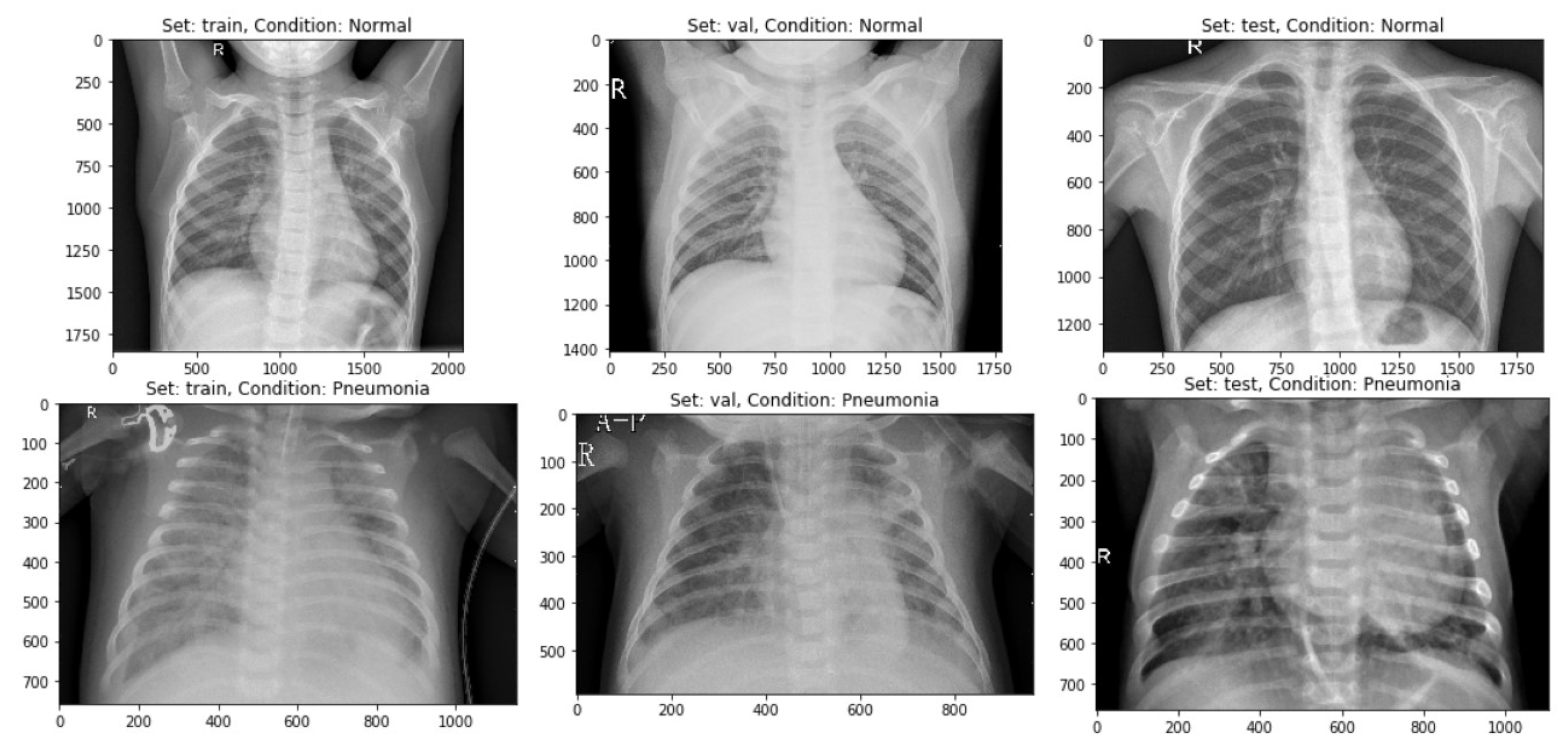
Sensors Free Full Text Detecting Pneumonia Using Convolutions And Dynamic Capsule Routing For Chest X Ray Images Html

Pin By Eve Emmanuelle Roy Hebert On T I M Diagnostic Imaging Med Student Medical Field

Pediatric Chest Supine View Radiology Reference Article Radiopaedia Org

Saber Sheath Trachea Radiology Case Radiopaedia Org Radiology Thoracic Trachea
2

Chest X Ray Of A 6 Month Old Child With An Icd The Active Can Is Download Scientific Diagram
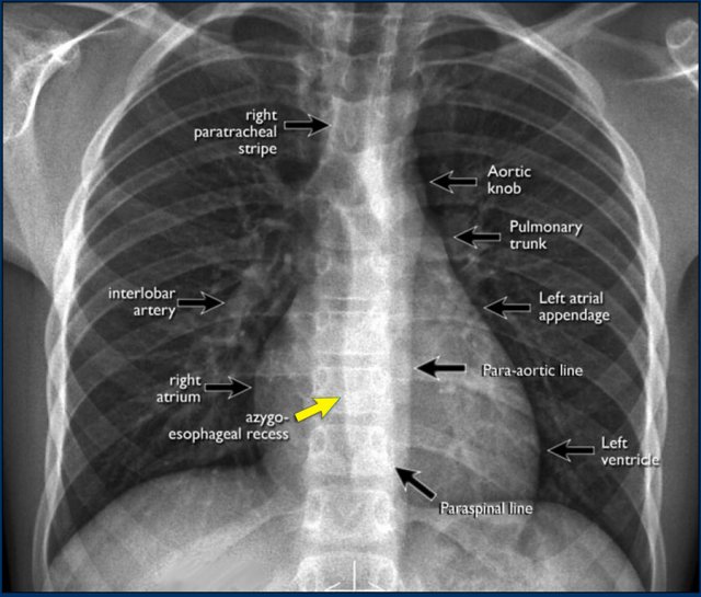
The Radiology Assistant Chest X Ray Basic Interpretation

Pediatric Chest Horizontal Beam Lateral View Radiology Reference Article Radiopaedia Org

Neonatal Radiography Part 1 Nomal Findings And The Basics Youtube